Final diagnosis:
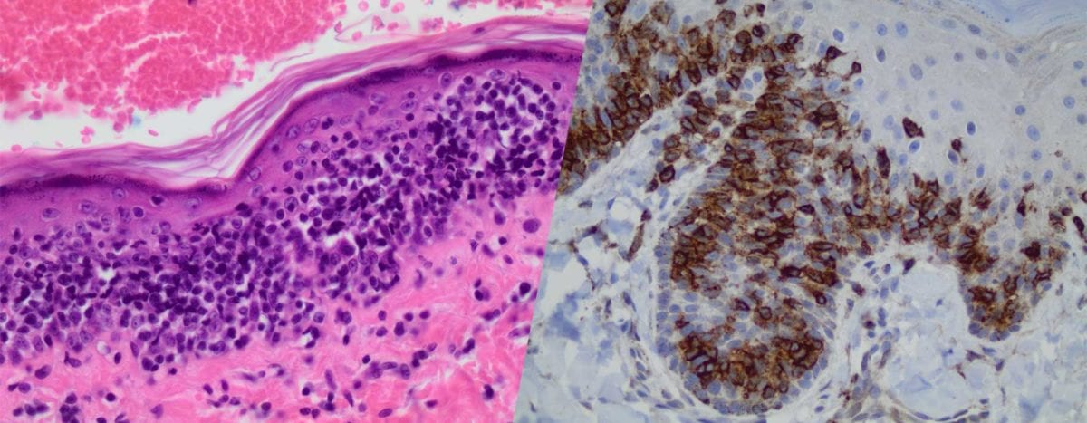
Epitheliotropic T-cell lymphoma
Discussion:
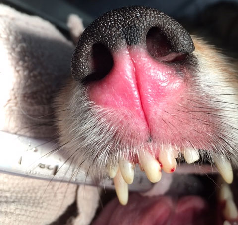
Epitheliotropic cutaneous lymphoma is an uncommon cutaneous neoplasm of older dogs. The disease has a heterogeneous clinical presentation that may include generalized erythema and scaling dermatitis, focal to multifocal erythema and/or ulceration, and nodular disease. Although the disease may present as diffuse dermatitis, lesions affecting mucocutaneous junctions and the oral cavity may occur alone or in concert with generalized dermatitis. Pruritus, sometimes severe, may occur and mimic allergic dermatitis. Lymphadenopathy may also be apparent. The diverse clinical presentation of this disease may present a clinical challenge, and surgical biopsy is recommended for definitive diagnosis.
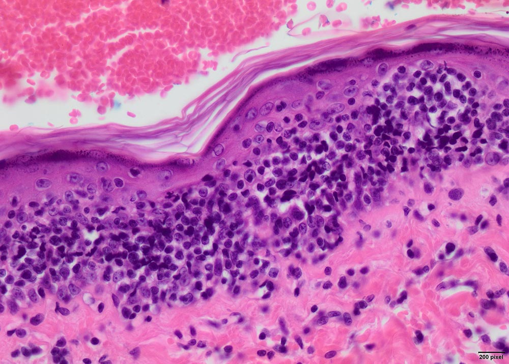
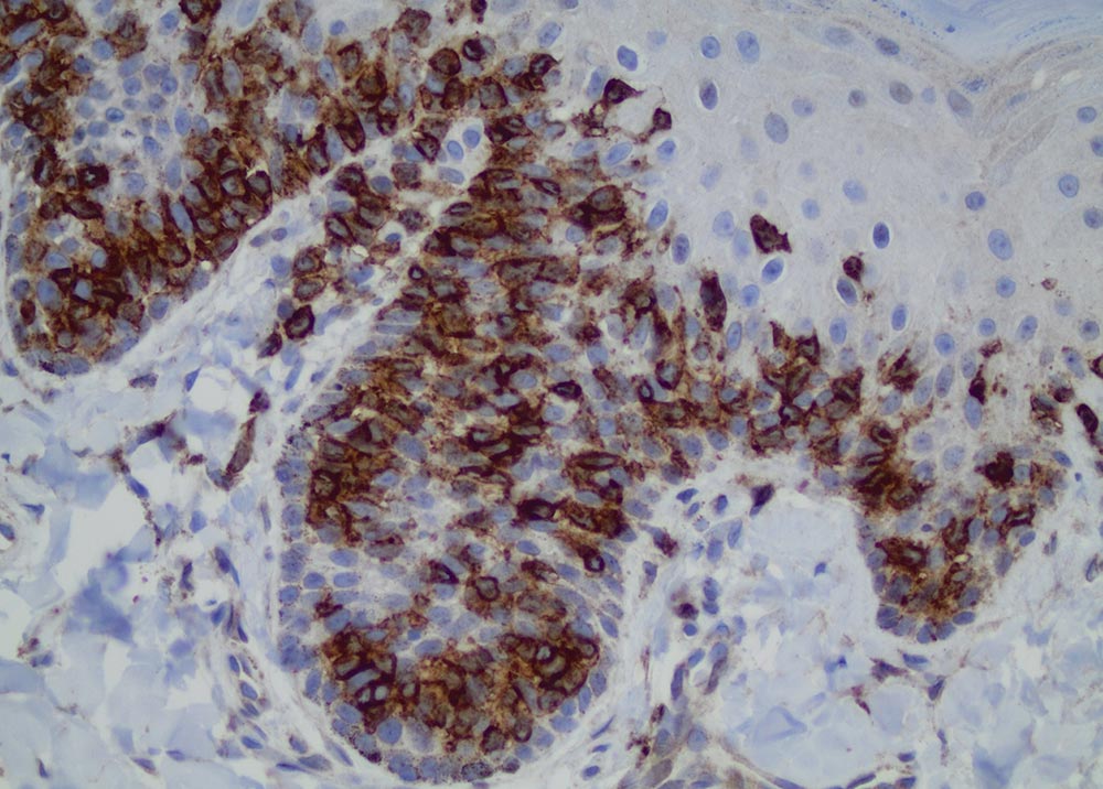
Microscopically, the diagnosis of epitheliotropic lymphoma is typically straightforward and characterized by the infiltration of the epithelium of the epidermis and adnexa by a uniform population of lymphocytes. However, in some cases lesions may overlap with those of other dermatoses and immunohistochemistry. Clonality testing (i.e. PARR testing) may be necessary to achieve a definitive diagnosis. In dogs, the vast majority of epitheliotropic cutaneous lymphomas are of T cell origin.
The prognosis for canine epitheliotropic cutaneous lymphoma is poor, with a reported median survival of six months. Therapies include chemotherapy, radiotherapy, and small molecule inhibitors although these have typically been utilized with little uniform long-term success.
Discoid lupus erythematosus (DLE) and uveodermatologic syndrome (UDS) were clinical differential diagnoses ruled out based on histopathology. However, DLE and UDS often present with similar clinical lesions as in this case including depigmentation, erythema, and/or crusting or ulceration of the nasal planum.
DLE is an immune-mediated condition of dogs targeting the dermal-epidermal junction that typically affects the face (nasal planum and periocular skin), although generalized and mucocutaneous forms are also described. The disease is exacerbated by UV light and German shepherd dogs are reportedly predisposed to multiple forms of lupus erythematosus but DLE may be observed in any breed. Diagnosis is typically via clinical history and biopsy. Histologic features in chronic cases of DLE may overlap with those of mucocutaneous pyoderma. Culture and clinical response to therapy may be necessary to help discern DLE from mucocutaneous pyoderma.
Uveodermatologic syndrome (UDS; Vogt-Koyanagi-Harada-like disease) is an immune-mediated disease of dogs presumably targeting melanocyte proteins resulting in a depigmenting dermatitis and uveitis. The nasal planum is often severely affected. Akitas are an over-represented breed for UDS, and uveitis, often occurring prior to or concurrent with dermatitis, may be severe and lead to blindness.
The distinction of these differential diagnoses can be clinically difficult and submitting biopsies to a deramatopathologist may help obtain the most accurate diagnosis which will help guide discussions of prognosis as well as therapeutics.
References:
- Rook KA. Canine and Feline Cutaneous Epitheliotropic Lymphoma and Cutaneous Lymphocytosis. Veterinary Clinics of North America: Small Animal Practice 2019;49:67–81.
- Fontaine J, Heimann M, Day MJ. Canine cutaneous epitheliotropic T-cell lymphoma: a review of 30 cases. Vet Dermatol 2010;21:267–75.
- Olivry T, Linder KE, Banovic F. Cutaneous lupus erythematosus in dogs: a comprehensive review. BMC Vet Res 2018;14:1–18.